

Presented by
Setareh Lavasani
DDS, MS, FGDIA
View Bio
Course Description
Oral Diagnosis, Oral Medicine, Oral Pathology, Radiology
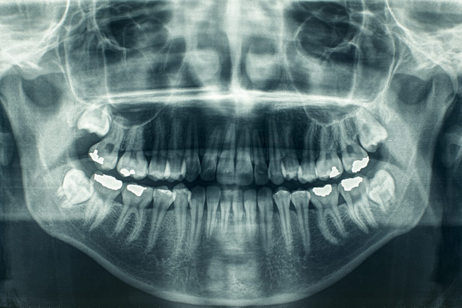
In this lecture, we will discuss how to develop an analytical strategy to review all 2D and 3D images systematically, and we will learn the importance of pattern recognition in diagnosing and differentiating benign and malignant lesions. We will also review common but important soft tissue calcifications in plain film and CBCT. We will also get an overview of paranasal sinuses and TMJ evaluation. Lastly, we will review the role of interprofessional communication in improving patient outcomes and the importance of selection criteria in using different imaging modalities for a specific task.
Course Objectives
At the completion of this course the participants should be able to:
- Review the most common radiographic presentations of malignant lesions in 2D and 3D.
- Develop an analytic strategy to evaluate each radiographic abnormality.
- Highlight the value of 3D CBCT in assessing head and neck abnormalities.
- Discuss best practices regarding following up on radiographic abnormalities from the need to watch and wait, treat, biopsy, and specialist referral.
- Emphasize the role of interprofessional communication in improved patient outcomes.

